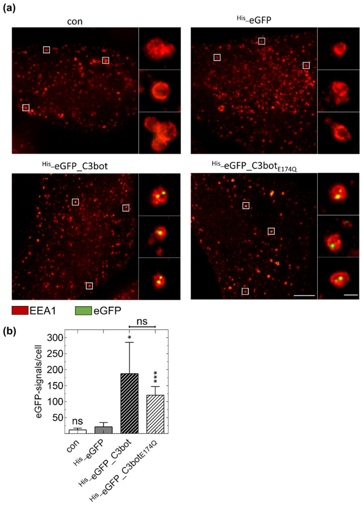
Figure 6.
Specific internalization of eGFP (green)-labeled C3bot and C3botE174Q into early endosomes (red) of mature human monocyte-derived DCs ex vivo. (a) Isolated human monocytes were differentiated into mature DCs. DCs were treated with 250 nM His_eGFP_C3bot, His_eGFP_C3botE174Q, His_eGFP for 30 min at 37 °C, or left untreated (con). After subsequent immunostaining, STED super-resolution microscopic images were captured. Magnified areas of each image are marked with white squares. The experiment was repeated with DCs differentiated from monocytes of five individual and independent donors. Scale bar corresponds to 5 µm (0.5 µm for the magnifications) and holds for all images. (b) The detected green spots were quantified for each treatment (n = 5 donors). Significant differences compared to the His_eGFP samples were tested using a Student’s t test (ns = not significant, * p < 0.05, *** p < 0.001). Comparing the samples with His_eGFP_C3bot and His_eGFP_C3botE174Q no significant differences were found as indicated above the graph.

