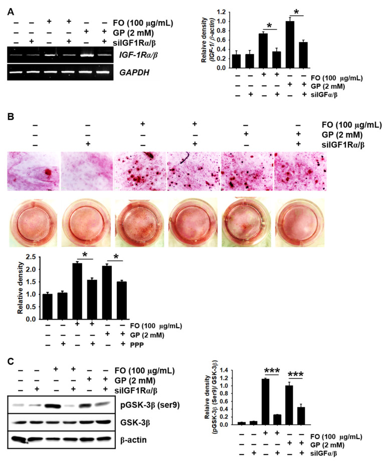
Figure 6.
Transient knockdown of IGF-1Rαβ reduces calcium deposition concomitantly with a decrease in phosphorylated GSK-3β at Ser9. MC3T3-E1 cells were seeded at a density of 2000 cells/cm2 and transfected with silencing RNA for IGF-1Rαβ (siIGF-1Rαβ) 48 h before stimulation with 100 µg/mL FO or 2 mM GP. (A) Total mRNA was extracted, and reverse transcription polymerase chain reaction was performed. GAPDH was used as an internal control. (B) After 7 days, the cells were stained with alizarin red for calcium deposition, and images were captured. (C) Three days after treatment with FO, total proteins were isolated and Western blotting was performed to measure the protein level of phosphorylated GSK-3β at Ser9 (left). Total GSK-3β and β-actin were used as internal controls. The expression of IGF-1Rαβ and phosphorylated GSK-3β at Ser9 relative to GAPDH and total GSK-3β levels is illustrated (right). Significant differences among the groups were determined using one-way ANOVA followed by Bonferroni correction. All data are presented as mean ± SEM (* p < 0.05 and *** p < 0.001 vs. untreated MC3T3-E1 cells). FO, fermented oyster (C. gigas) extract.

