Figure 1.
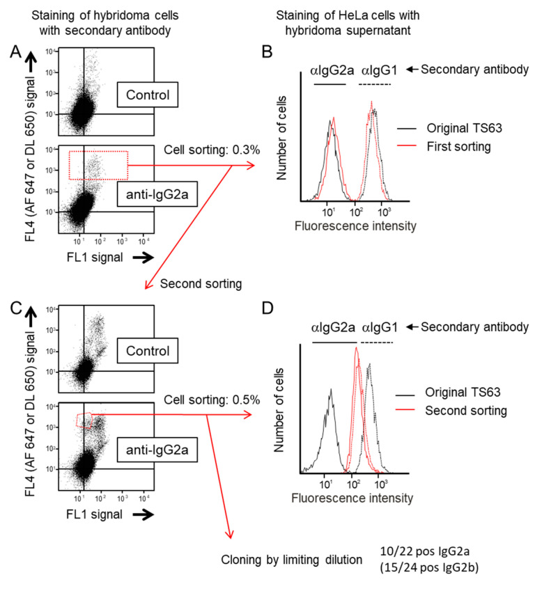
Selection of IgG2a variants of TS63. (A) Hybridoma cells were labelled with either a control secondary antibody (a goat anti-rabbit coupled to Dylight 650 (DL 650)) or a goat anti-mouse IgG2a coupled to Alexa Fluor 647 (AF 647). The dot plots show for each cell the value of the Dylight 650 or Alexa 647 fluorescence in the far red Channel (FL4) and that of the autofluorescence in the green channel (FL1). Note that there is no difference between the two labelings indicating that most of the “positive” signal is non-specific. The gate used to sort the cells with the highest level of staining is drawn in the bottom dot-blot. (B) After being grown for a few days, the supernatant of the sorted cells was used to stain HeLa cells by indirect immunofluorescence, using either anti (α)-IgG1 or anti-IgG2a polyclonal antibodies coupled to Alexa 647 as secondary reagents. A staining with the conditioned medium of parental TS63 cells was performed in parallel. The fluorescence staining of the cells was analyzed by flow-cytometry. Note that the supernatant of sorted cells stains HeLa cells slightly better than that of parental cells when the binding of the mAb is revealed by an anti-IgG2a antibody. (C) After amplification, the cells sorted in A were subjected to a second sorting. Note that there is a specific cell population uniquely detected by the anti-mouse IgG2a labeling. The gate used to sort the cells is drawn in the bottom dot-blot. (D) Labelling of HeLa cells as in B after growing the cells sorted the second time for a few days. Note that the staining of HeLa cells by the supernatant of the sorted cells is similar whether the binding of the mAb is revealed by an anti-IgG2a antibody or an anti-IgG1 antibody.

