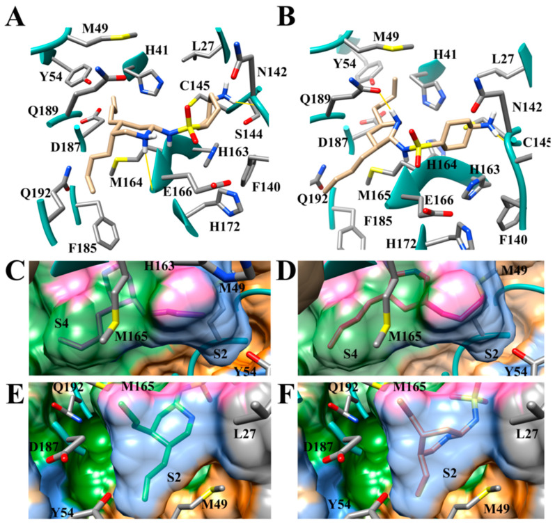
Figure 12.
Modes of binding of SFY derivatives. (A) SD34 (tan); (B) LEA4 (tan); (C) SD34 (purple); (D) LEA4 (pink); (E) SD35 (light green); (F) SD36 (brown). In panels (A) and (B) it can be seen that the aliphatic tails bind in the S2 and the S4 subsites. In panels (C) through (F) of the surface of the subsites, each of the bound aliphatic substituents is shown. The S2 subsite is colored blue, and the S4 subsite is green. Grey regions are portions of the S1′ subsite. Hydrogen bonds are indicated by solid yellow lines.

