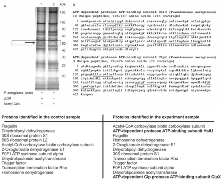
Figure A6.
Rac substrate profiling. (A) Immunoaffinity enrichment of the Rac substrates from P. aeruginosa cell lysate. The arrow indicates the additional acetylated protein in the presence of recombinant Rac and acetyl-CoA (lane 1), compared to the control sample (lane 2). (B) MS analysis reveals that HslU and ClpX are present in lane 1 but not in lane 2, with sequence coverage of 24% and 7%, respectively. The identified peptide sequences are underlined.

