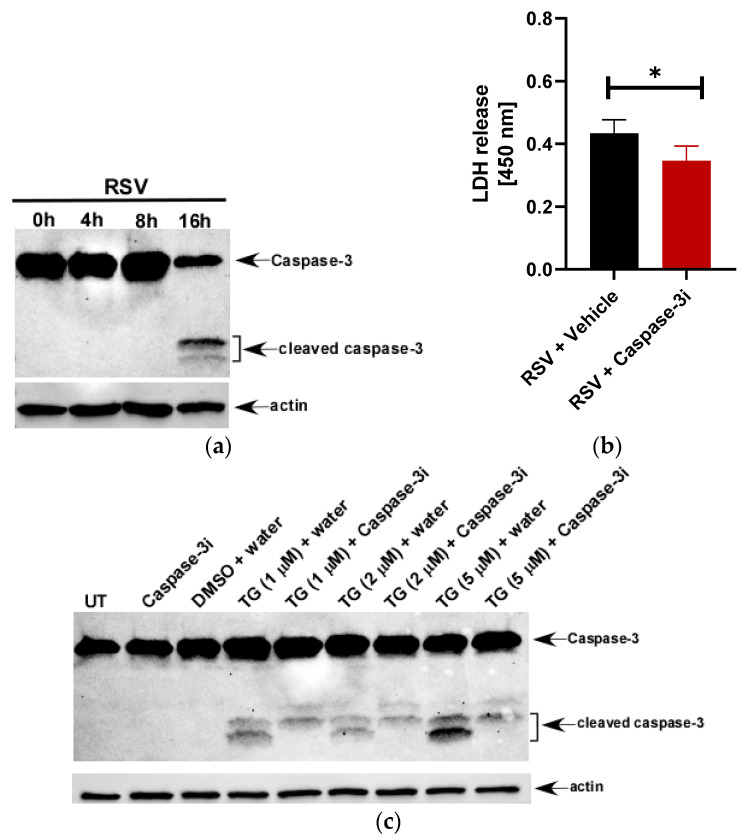
Figure 2.
LDH release during RSV infection of macrophages is primarily due to lytic cell death. (a) Human THP-1 macrophages infected with RSV (MOI = 1) for 0 h–16 h were subjected to Western blotting with anti-caspase-3 antibody that can detect both the full length and cleaved fragments of activated caspase-3. (b) Human THP-1 macrophages were infected with RSV (MOI = 1) in the presence of either vehicle (water) or the caspase-3 inhibitor Ac-DEVD-CHO (100 µM). LDH release was measured (at OD of 450 nm) at 16 h post-infection infection (n = 16 technical replicates from two independent experiments). * p ≤ 0.05 using a Student’s t-test. (c) Untreated (UT) and THP-1 cells treated with indicated concentrations of thapsigargin (TG) in the presence of either water (vehicle control) or caspase-3 inhibitor Ac-DEVD-CHO (100 µM) for 48 h were subjected to Western blotting with anti-caspase-3 antibody that can detect both the full length and cleaved fragments of activated caspase-3. TG is soluble in DMSO and therefore, a lane for DMSO + water was included for the Western blot analysis. Western blot data is representative of two-three independent experiments with similar results. Caspase-3i: caspase-3 inhibitor Ac-DEVD-CHO.

