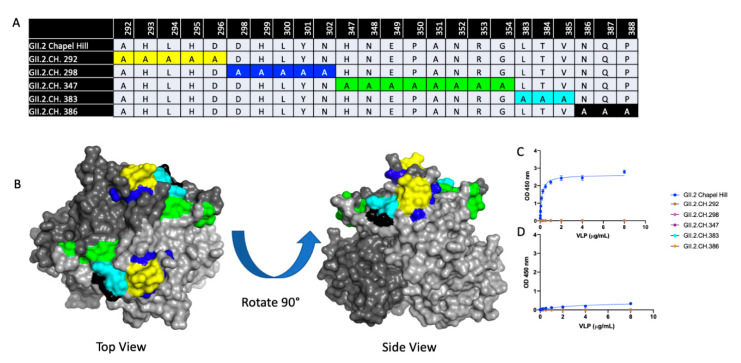
Figure 5.
Characterization of GII.2 Chapel Hill Alanine mutant VLPs. (A) A panel of 5 alanine mutant VLPs were constructed comprising variable residues between GII.2 1976 SMV and GII.2 Chapel Hill within the P2 domain. Sequence alignments of variable residues within the 5 alanine mutants are compared against the Chapel Hill backbone. Color coordinated highlighted regions are highlighted on the GII.2 P domain dimer in (B). VLP binding to A and B saliva (C,D) were analyzed via one-site specific binding curve, fitted with error bars representing standard error of the mean.

