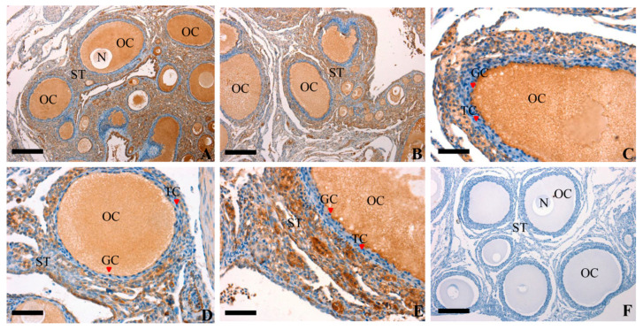
Figure 2.
Representative photomicrographs of RAC1 protein immunolocalization (in brown) in the prehierarchical follicle of ovarian. Panel (A,B), oocytes and GCs were strongly stained within the different size of prehierarchical follicles; Panel (C,D), a larger developing prehierarchical follicles, which contains two or three layers of GCs; Panel (E), a larger prehierarchical follicles and more layers of GCs and thecal cells developing in this stage; Panel (F), RAC1 negative controls, which were treated with pre-immune serum and no specific staining was observed. Oocyte (OC), granulosa cell (GC), theca cell (TC), stroma (ST) and nucleus (N) are indicated. Scale bar = 200 μm (A,B,F); 100 μm (C,D,E).

