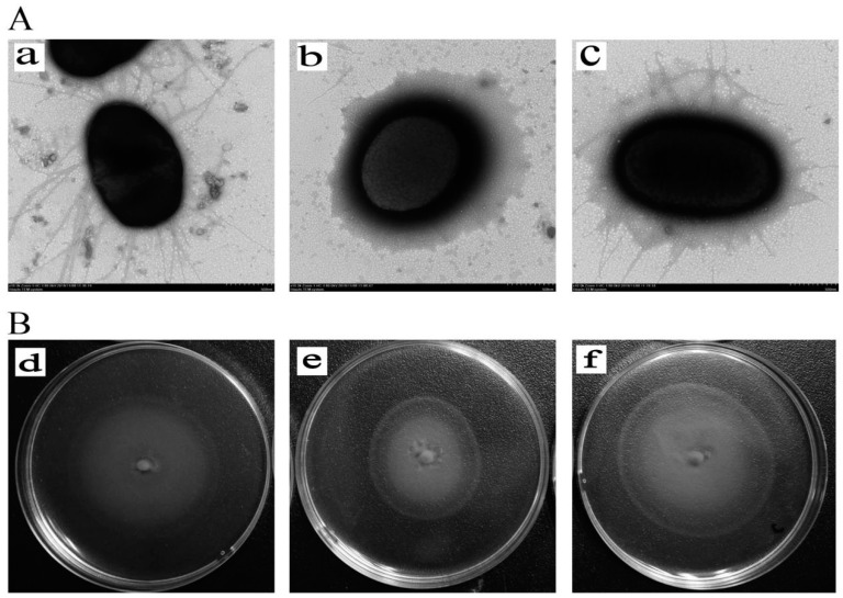
Figure 3.
(A). The flagella of AE81, AE81ΔyqeI and AE81ΔyqeI-pCmyqeI in the transmission electron micrographs views (×10,000). (a) The morphological observation of AE81 (×10,000); (b) the morphological observation of AE81ΔyqeI (×10,000); (c) the morphological observation of AE81ΔyqeI-pCmyqeI (×10,000) (B). The observation of motility ability. (d) The motility circle of the wild strain AE81; (e) the motility circle of AE81ΔyqeI; (f) the motility circle of AE81ΔyqeI-pCmyqeI.

