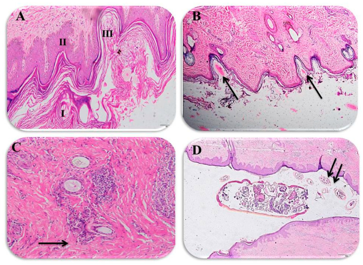
Figure 3.
Histopathological findings for the infected camels: (A) the epidermis is thickened by compact hyperkeratosis (I) and acanthosis (II), which are in response to both the mite itself and also to the self-trauma caused by intense pruritus, and also shows the burrowing tunnels of the mites (III), ×100 magnification; (B) histological section showing Sarcoptic scabiei eggs in tunnels in the epidermis of an infested camel (arrow), ×200; (C) dermis showing inflammatory cell infiltration around the blood vessels and perifolliculitis (arrow), ×100; and (D) adult Sarcoptic scabiei and its eggs within the migrating tunnels of the camel skin (arrow), ×200.

