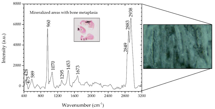
Figure 5.
Raman spectra of mineralized areas of bone metaplasia of the mixed carcinoma (sample #3) and, on the right side, the corresponding microscopic image (50×, Raman spectrometer system). In the inset is the microscopic image (2.5×) of a section of sample #3 where mineralized areas are stained black by means of the Von Kossa-Van Gieson modified method.

