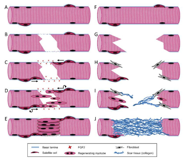
Figure 3.
Skeletal muscle regeneration. Differences in regeneration following small scale skeletal muscle injuries (A–E) vs. volumetric muscle loss (F–J). Healthy muscle tissue (A) incurs a small-scale injury, which damages the myofiber and its surrounding basal lamina (B). The disrupted basal lamina releases sequestered growth factors including fibroblast growth factor 2 (FGF2) and satellite cells are activated, proliferating and migrating into the wound site along the basal lamina (C). Satellite cells begin fusing to form myotubes while simultaneously self-renewing (D). Resulting tissue is fully recovered, with aligned myotubes (E). Healthy muscle tissue (F) incurs a large-scale VML injury, which destroys the majority of native basal lamina and satellite cells (G). Without these cues, satellite cell-mediated regeneration is diminished, and fibroblasts begin infiltrating the wound (H). The injury is characterized by sparse and misaligned myoblast infiltration and collagen deposition (I), resulting in scar tissue formation and decrease in muscle function (J).

