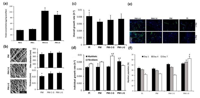
Figure 2.
Comparison of the cell growth of the fibroblasts and myoblasts on each type of the nanofiber scaffolds. Abbreviations: Pl, PM, PM-C, PM-L, PM-C-G, and PM-L-G are the plastic surface, PMMA, PMMA-collagen, PMMA-laminin, PMMA-collagen-genipin, and PMMA-laminin-genipin. (a) Protein concentration. Genipin aided in collagen and laminin adsorption to nanofibers. (b) SEM image, diameter, and thickness of the electrospun nanofiber. (c,d) Overall and individual cell growth rate, respectively. PM-L-G enhanced the growth of myoblasts while it suppressed that of fibroblasts. (e) Fluorescence microscopy showing the myoblasts (green) and overall nucleus (blue). (f) Myoblast population within 7 days. Only PM-L-G showed a growing population of myoblasts. Reproduced from [24], copyright open access by MDPI 2017.

