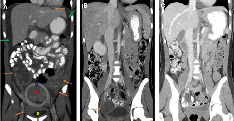
Figure 3.
Coronal contrast-enhanced computed tomographic (CT) images of the abdomen and pelvis at presentation and after treatment. (A) At presentation demonstrating presence of multiple abdominal/pelvic tumors (orange arrows), intrauterine mass (red X), and malignant ascites (green arrows). (U) Uterus, (B) bladder. (B) After six cycles of chemotherapy demonstrating treatment response with resolution of malignant ascites and significant decrease in tumor burden with residual right pelvic tumor (orange arrow). (C) After surgical resection and completion of chemotherapy demonstrating complete treatment response with no detectable tumor.

