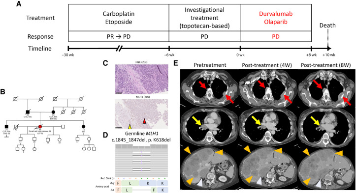
Figure 1.
Patient clinical course and germline mismatch repair (MMR) deficiency. (A) Patient clinical course, treatment, response, and timelines. (B) Patient genogram. (C) Hematoxylin and eosin staining (top) and MLH1 IHC (bottom). The red arrowhead in the bottom panel indicates loss of MLH1 expression on tumor cells, whereas the yellow arrowhead indicates intact MLH1 expression in normal hepatic cells. Black bars, 100 µm. (D) Integrated Genomics Viewer panel of germline MLH1 mutation. (E) Contrast-enhanced computed tomography (CT) scans pretreatment (left), 4 wk (middle) after treatment, and 8 wk (right) after treatment. Red arrows, yellow arrows, orange arrowheads, and light blue arrowheads indicate pleural lesions, right mediastinal lymph nodes, liver lesions, and right adrenal mass, respectively. (dMMR) Mismatch repair deficiency, (W) week, (PR) partial response, (PD) progressive disease, (COL) colorectal cancer, (BR) breast cancer, (THY) thyroid cancer, (UT) endometrial cancer, (H&E) hematoxylin and eosin, (IHC) immunohistochemistry staining, (Ref) reference, (Alt) alteration.

