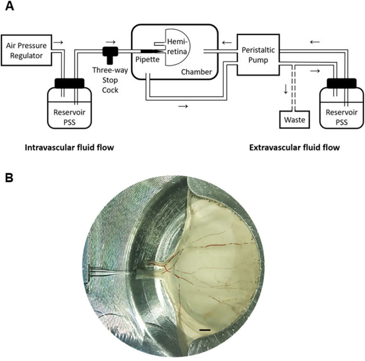
Figure 1.
The experimental setup. (A) The perfusion system. Arrows indicate the direction of flow in, respectively, the intravascular (left) and the extravascular (right) perfusion systems. (B) An upper porcine hemiretina mounted in the tissue chamber. Stagnant erythrocytes are seen in the larger vessels. Bar = 2 mm.

