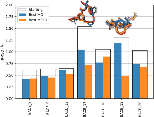
Fig. 9.
Comparison of MELD refinement results, MD refinement results, and top-1 starting conformations. The best MELD refined pose, best MD refined pose, and top-1 starting pose are shown in orange bars, blue bars, and white boxes, respectively. The corresponding structures for BACE_17 and BACE_19 are shown above the bars in the same colors; the native structure is shown in red

