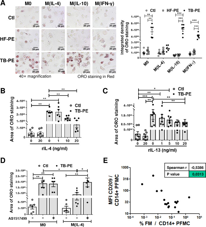
Fig 1. The IL-4/STAT6 Axis prevents the formation of foamy macrophages.
Human macrophages were left untreated (M0) or polarized with either IL-4 (M(IL-4)), IL-10 (M(IL-10)) or IFN-γ (M(IFN-γ)) for 48 h, treated or not with the acellular fraction of TB pleural effusions (TB-PE) or heart-failure-associated effusions (HF-PE) for 24 h and then stained with Oil Red O (ORO). (A) Left panel: Representative images (40× magnification), right panel: the integrated density of ORO staining. (B-C) Quantification of area of ORO staining of macrophages polarized with either different doses of recombinant IL-4 (B) or IL-13 (C) for 48 h and exposed to TB-PE for further 24 h. (D) Quantification of area of ORO staining of M0 and M(IL-4) macrophages treated with TB-PE and exposed or not to AS1517499, a chemical inhibitor of STAT6. Values are expressed as means ± SEM of six independent experiments, considering five microphotographs per experiment. (E) Correlation study between the mean fluorescence intensity (MFI) of CD209 cell-surface expression in CD14+ cells from TB pleural cavity and the percentage of lipid-laden CD14+ cells within the pleural fluids mononuclear cells (PFMC) (n = 16) found in individual preparations of TB-PE. Spearman’s rank test. Friedman test followed by Dunn’s Multiple Comparison Test: *p<0.05; **p<0.01; ***p<0.001 as depicted by lines.

