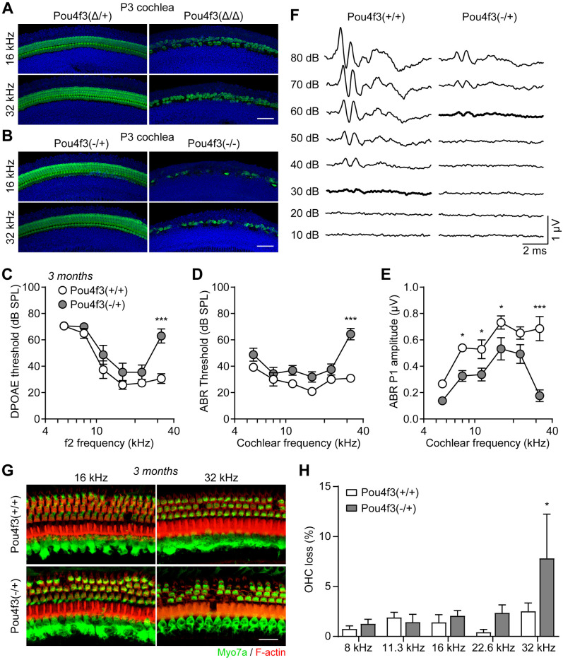
Fig 3. Heterozygous Pou4f3 knockout mice display high frequency hearing loss and degeneration of outer hair cells.
(A-B) Myo7a immunostaining of cochlear hair cells from (A) P3 Pou4f3(Δ/+) and Pou4f3(Δ/Δ) mice and (B) P3 Pou4f3(-/+) and Pou4f3(-/-) mice. Scale bars were 50 μm. (C) DPOAE threshold, (D) ABR threshold and (E) ABR P1 amplitudes of 3 months old Pou4f3(+/+) and Pou4f3(-/+) mice. * P < 0.05 and *** P < 0.001 by two-way ANOVA, n = 6–8 mice of each genotype. (F) ABR waveforms of 3 months old Pou4f3(+/+) and Pou4f3(-/+) mice at 32 kHz. Data were mean of n = 6–8 animals. ABR thresholds of each genotype were highlighted. (G) Myo7a immunostaining images of the cochlear sensory epithelia from 3 months old Pou4f3(+/+) and Pou4f3(-/+) mice. Hair cells and F-actin was labelled with Myo7a (green) and Rhodamine-phalloidin (red), respectively. Scale bar was 20 μm. (H) Percentage of outer hair cell loss in Pou4f3(+/+) and Pou4f3(-/+) mice at various cochlear frequencies. * P < 0.05 by two-way ANOVA, n = 8 cochleae of each genotype.

