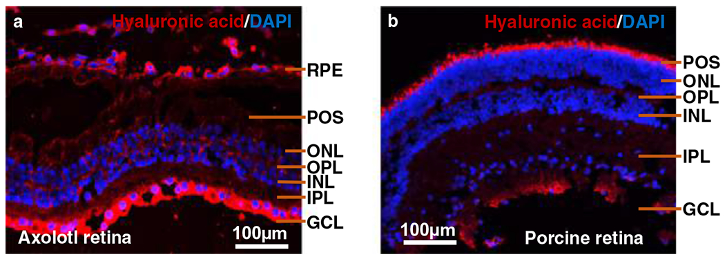
Fig. 1.

Cross-sectional anatomy of the (a) Axolotl and (b) porcine retina. Immunohistochemistry was performed to visualize expression of hyaluronic acid in the axolotl and porcine retina. Primary antibody (anti-hyaluronic acid sheep polyclonal, 1:1000 in goat serum block, Abcam) and secondary antibody (1:400 in PBS, Alexa FluorTM 594 donkey anti-sheep IgG, Invitrogen) were used to stain HA red while DAPI used to stain the nucleus blue. POS: Photoreceptor outer segments; ONL: outer nuclear layer; OPL: outer plexiform layer; INL: Inner nuclear layer; IPL: Inner plexiform layer; GCL: Ganglionic cell layer; RPE: Retinal pigment epithelium
