. 2020 Oct 13;9:e60581. doi: 10.7554/eLife.60581

Figure 6. Interactions stabilizing the priming of the PfMyoA PPS state.

(a) Overall structure of PfMyoA•FL-PPS. The priming of the lever arm is stabilized by interactions between elements of the lever arm and the motor domain (boxed in black). (b) Zoom on the lever arm/motor domain interface. The residues involved in the interaction are labeled. Key residues that have been mutated to disrupt the interface (see d) are labeled red. (c,d) MD simulations comparing the WT and triple mutant R707A/E711A/Y714A. (c) The primed PPS state is stable during the entire duration of the simulation (320 ns). (d) In contrast, the priming is lost with the triple R707A/E711A/Y714A mutant and the position of the lever arm deviates by up to 54°.

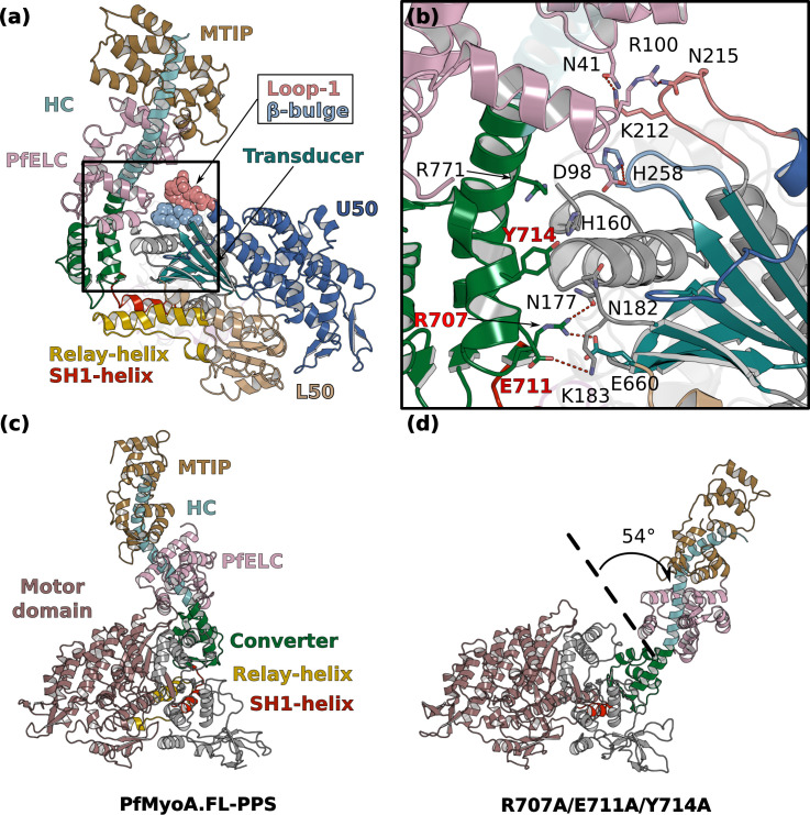
Figure 6.

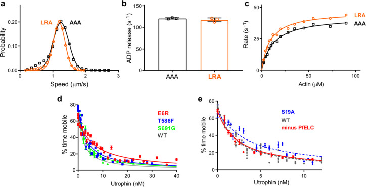
Figure 6—figure supplement 1. Transient kinetics and genetic engineering of the parasite.

Figure 6—figure supplement 1.

(a, e) Triple mutants that affect the atypical priming of PfMyoA. speed, ADP-release rate, and actin-activated ATPases for LRA (see also data presented in Figure 2) compared with the AAA mutant (LRA: R707L/E711R/Y714A; AAA: R707A/E711A/Y714A).(a) Speed distributions from a representative in vitro motility assay for LRA (1.19 ± 0.18 μm/s, n = 4092) and AAA (1.24 ± 0.24 μm/s, n = 3030). (b) ADP-release rates from acto-PfMyoA for LRA (117 ± 4 s−1) and AAA (122 ± 2 s−1). three experiments, one protein preparation of AAA. (c) Actin-activated ATPase activity for LRA (Vmax = 46.8 ± 1.0 s−1; Km = 9.1 ± 0.6 μM) and AAA (Vmax = 42.7 ± 0.9 s−1; Km = 12.3 ± 0.8 μM). Error, SE of the fit. two experiments, one protein preparation of AAA. (d) Ensemble force measurements using a utrophin-based loaded in vitro motility assay, showing more data and an expanded x-axis compared with the main Figure 2f. Temperature, 30°C. (e) Ensemble force measurement of FL PfMyoA with only MTIP light chain bound. A myosin that produces more force requires higher utrophin concentrations to slow motion. Minus PfELC: 1.57 ± 0.18 nM (red diamonds and solid red line fit); WT, 1.40 ± 0.08 nM; S19A, 2.42 ± 0.17 nM. WT and S19A data are from Frénal et al., 2017. Error, SE of the fit. Data for minus PfELC are from one protein preparation and two experiments. Skeletal actin was used for all experiments. Temperature, 30°C.
Figure 6—figure supplement 1—source data 1. Source data for kinetic experiments presented in Figure 6—figure supplement 1.
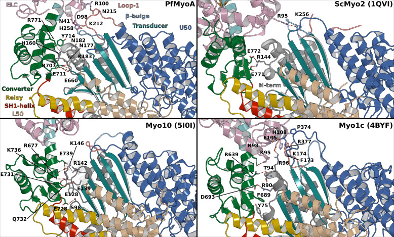
Figure 6—figure supplement 2. Interactions between the motor domain (MD) and the lever arm stabilizing the priming in PPS in different myosins.

Figure 6—figure supplement 2.

Important interactions occur in PfMyoA between the converter and the N-terminal subdomain while the PfELC is involved in interactions with Loop1 and the β-bulge of the Transducer. In comparison, limited interactions occur in ScMyo2 consistent with smaller priming of the lever arm. Myo10 is another highly primed myosin but the interactions involve mainly the Converter with the N-terminal subdomain, without contribution from the Transducer. The priming of another highly primed PPS structure from Myo1c requires interaction of the CaM light chain with Loop1 and the HO-linker of the Transducer.