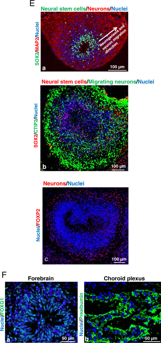
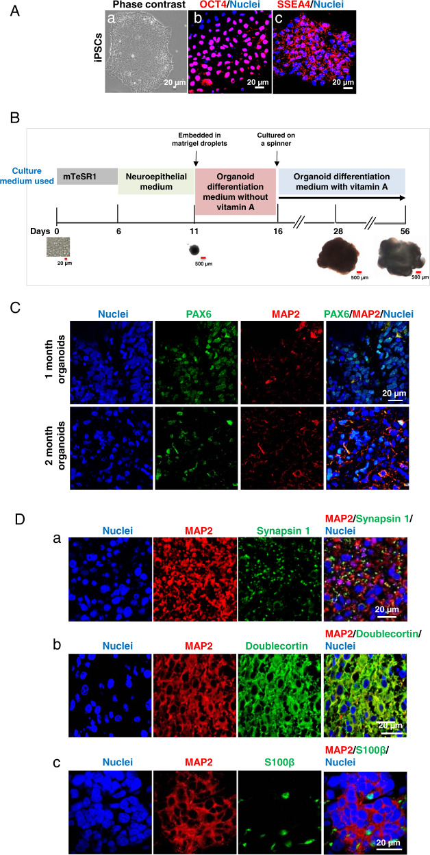
Fig. 1. Characterization of human-induced pluripotent stem cells (iPSCs) and iPSC-derived cerebral organoids over the two-month differentiation process from iPSCs to cerebral organoids.

A Human iPSCs grew as colonies in the culture and expressed pluripotent stem cell markers octamer binding transcription factor 4 (OCT4) and stage-specific embryonic antigen-4 (SSEA4). The phase-contrast image shows the colonies of iPSCs maintained in mTeSR1 stem cell culture medium (a). Confocal images of the cells with immunofluorescence staining demonstrate that iPSCs colonies expressed OCT4 (red) in the nuclei (b) and SSEA4 (red) in cell member (c). Nuclei were stained with Hoechst 33342 in blue. Scale bar = 20 µm. B The schematic depicts the procedure for the generation of organoids from iPSCs by the use of chemically defined medium and plating strategies. Singularized iPSCs cultured in stem cell medium mTeSR1 aggregated into embryoid bodies in ultra-low attachment plates (day 0 to day 6), begun to differentiate into neuroepithelial tissue, and resulted in the formation of cerebral organoids following Matrigel™ embedding at day 11. The organoids grow bigger over time in the culture. Scale bar = 20 or 500 μm. C Cerebral organoids develop over time in the culture. Immunofluorescent staining marked the expression of the paired box protein 6 (PAX6) in neural epithelial progenitor cells (green) in 1-month organoids, which was markedly reduced in 2-month organoids. Microtubule-associated protein 2 (MAP2)-positive neurons (red) were expressed in 1-month cerebral organoids, but more fluorescence signals were observed in 2-month organoids. Blue are cell nuclei. Scale bar = 20 µm. D Cerebral organoids contain neurons and astrocytes. In 2-month old, immunofluorescent staining for pre-synapse marker synapsin I (green; distributed in a punctuate pattern) shows a vast number of synapses between MAP2-positive neurons (red) (a). The neurons (red) were immature as evidenced by the expression of doublecortin (green) (b). Astrocytes, as indicated by the S100 calcium-binding protein β (S100β) expression (green) were also present with neurons (red) in the organoids (c). Nuclei were stained in blue. Scale bar = 20 μm. E Two-month cerebral organoids display well-organized elaborate cellular laminar organization and architectures (neural stem cells and neurons expressing different cortex layer neuron markers are located in the different layers of the organoid tissues). SOX2-positive neural stem cells (green) are located on the apical side and neurons (red) are located on the basal side (a), suggesting that neural stem cell-derived neurons migrate from the apical toward the basal side. The white arrow indicates the migration direction of differentiated neurons. Neurons in cerebral organoids also expressed deep-layer cortical neuron markers CTIP2 (b, green) and FOXP2 (c, red). Blue represents cell nuclei. Scale bar = 100 μm. F Cerebral organoids display FOXG2-positive (green, a) forebrain and prealbumin-positive choroid plexus-like (green, b) brain regions. Blue represents cell nuclei. Scale bar = 50 μm.

