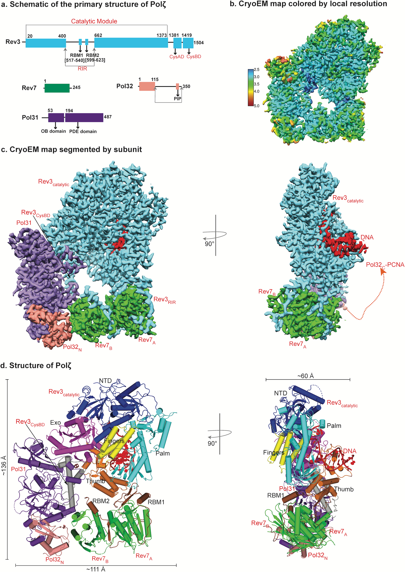
Fig. 1.

The architecture of DNA bound Polζ holoenzyme. a, Schematic of the primary structure of S. Cerevisiae Polζ subunits. Different colors denote each subunit. b, Near atomic resolution cryo-EM density map of DNA bound Polζ holoenzyme colored by local resolution. c, The three-dimensional reconstruction of Polζ holoenzyme viewed (left) perpendicular and parallel (right) to the DNA axis. A red dashed connector represents disordered Pol32c and the arrowhead marks the putative interaction location with PCNA. d, Cryo-EM structure of DNA bound Polζ colored by domain, and viewed from the same orientations as in (c).
