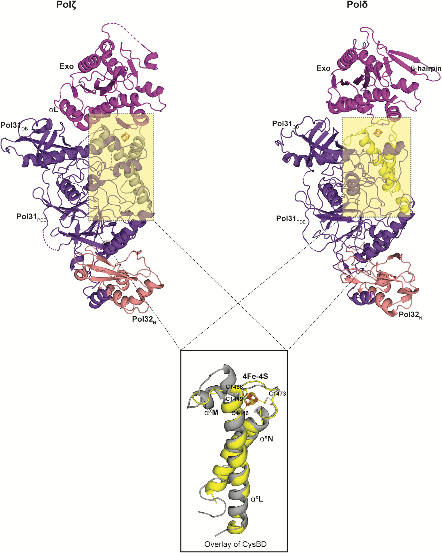
Extended Data Fig. 7. Comparison between the CysBD of Polζ and Polδ.

A superimposition of the CysBD of the Polζ (left; grey in color) and Polδ (right; yellow in color; PDB ID: 6P1H) shows conservation in its overall topology. Notably, helix αXM in Polζ CysBD has been substituted by a loop in Polδ (PDB ID: 6P1H). All the four cysteines interacting with the 4Fe-4S cluster in Rev3 are also highlighted..
