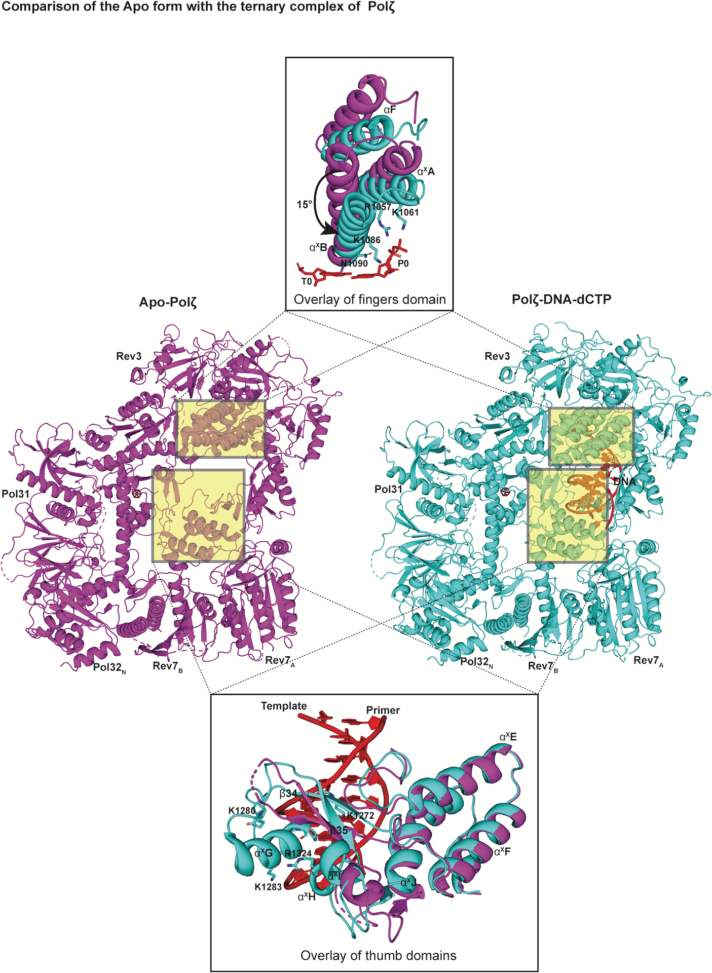
Fig. 6:

Conformational changes upon DNA binding. Structures of the ‘open’ apo state of Polζ (left, colored in magenta) in comparison to its ‘closed’ ternary complex (right, colored in cyan) viewed perpendicular to the DNA axis. An overlay of the fingers and the thumb domain of both states highlight significant conformational changes. In addition to the inward rotation of the fingers domain, various structural elements and loops (including αxG highlighted here) in the thumb domain become ordered upon DNA binding.
