
Fig. 4.
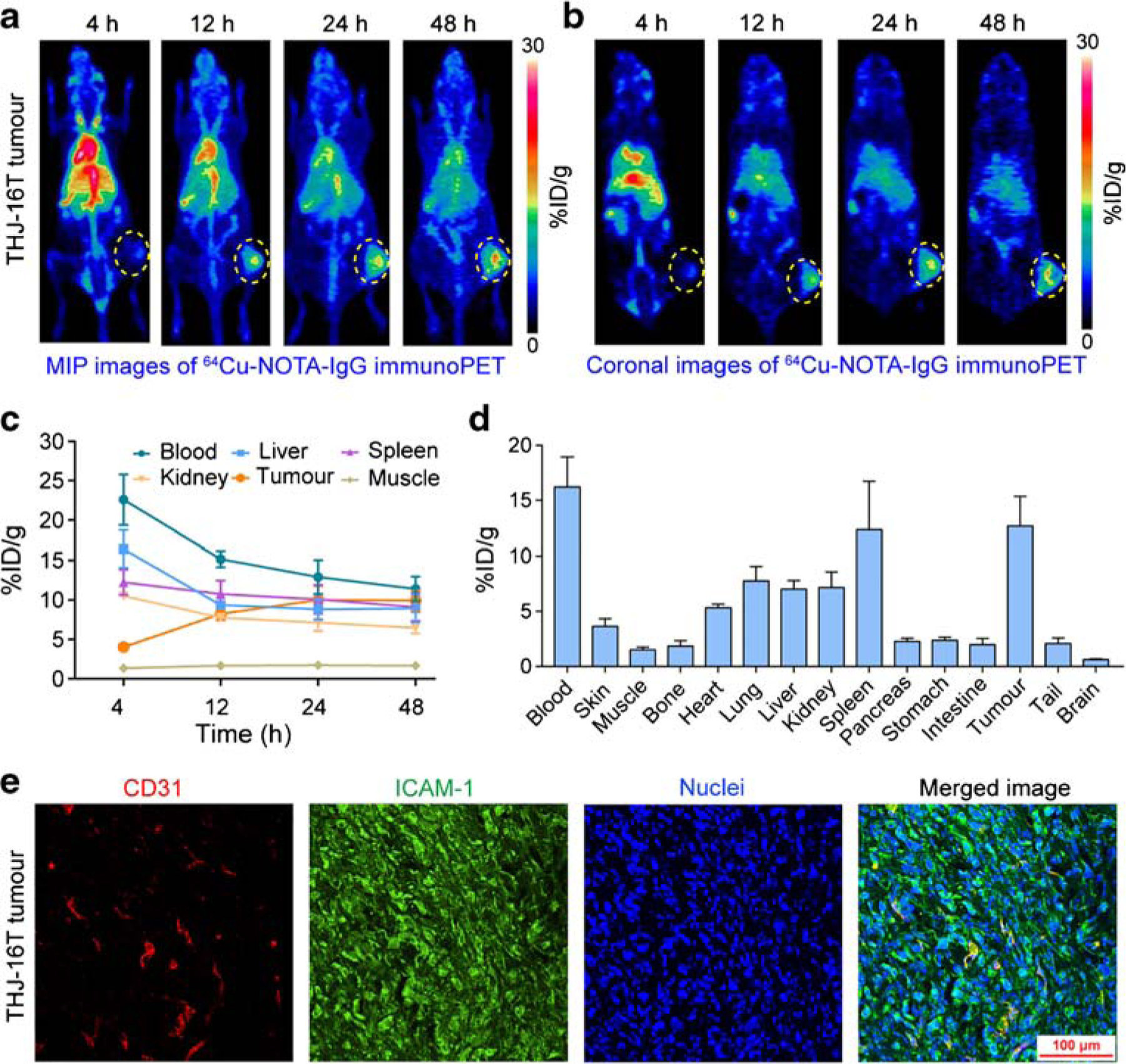
64Cu-NOTA-IgG immunoPET imaging of ICAM-1-positive subcutaneous anaplastic thyroid cancers. a, b 64Cu-NOTA-IgG immunoPET imaging of subcutaneous THJ-16T-bearing R2G2 mice. Maximum intensity projection (MIP) and coronal images of a representative mouse at different imaging time-points were given. c Region of interest analysis of 64Cu-NOTA-IgG immunoPET imaging data. d Biodistribution study at 48 h post-injection of 64Cu-NOTA-IgG. e Immunofluorescent imaging of a subcutaneous THJ-16T tumor. The tumor section was stained for CD31 (red), ICAM-1 (green), and nuclei (blue). The tumors were indicated by yellow dotted circles