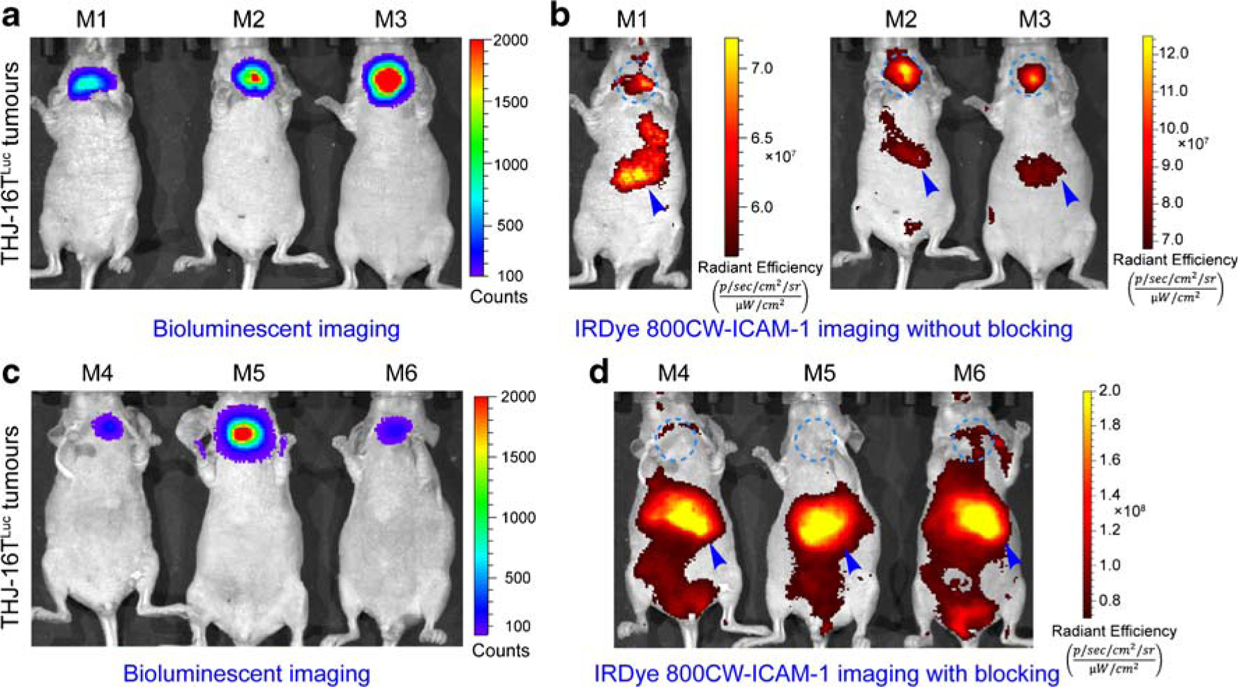
Fig. 6.

Bioluminescent imaging (BLI) and fluorescent imaging of orthotopic ATC models without or with R6–5-D6 blocking. a BLI of mice in the ICAM-1-targeted imaging group without R6–5-D6 blocking. The given image showed the growth of THJ-16TLuc tumors. b Fluorescent imaging of the same mice 48 h after injection of IRDye 800CW-ICAM-1. The fluorescent tracer was eliminated from the hepatobiliary system with a proportion deposited in the orthotopic THJ-16TLuc tumors. c BLI of mice in the blocking group showed comparable tumor burden. d Fluorescent imaging of the mice in the blocking group, which were injected first with a blocking dose of R6–5-D6 and then with the IRDye 800CW-ICAM-1. Fluorescent imaging acquired 48 h after injection of the tracer showed negligible signal in the thyroid areas, indicating saturation of the target by the blocking dose of R6–5-D6. The tumor areas were indicated by blue dotted circles and livers were indicated by blue arrowheads
