Figure 1:

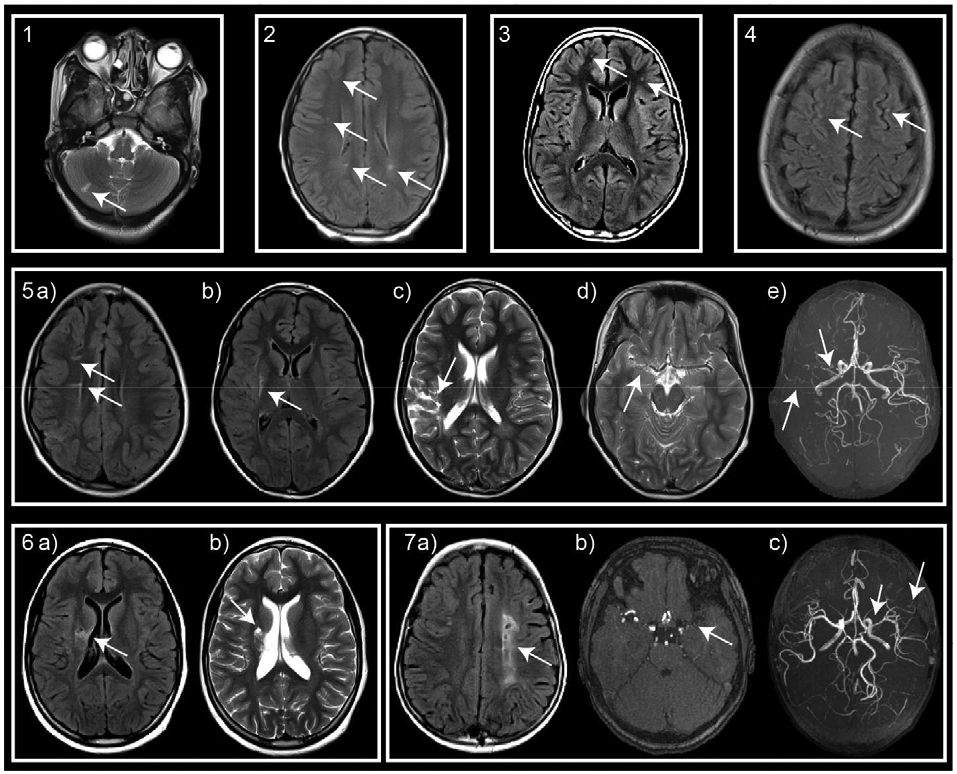
Neuroimaging evidence of CVD in HIV-infected patients. T2, T2 FLAIR, and reconstructed time of flight images demonstrate: participant 1: cerebellar infarct; participants 2-4: small foci of hyperintensities; participant 5: small infarcts in the a) right corona radiata, b) right putamen, c) right MCA territory, and vessel abnormalities consisting of d) high grade narrowing of the right M1 segment. e) MRA confirms M1 stenosis and demonstrates a paucity of distal MCA flow signal and net-like collateralization; participant 6: infarcts in the right basal ganglia and corona radiata; and participant 7: a) extensive infarct in the left centrum semiovale, b) left supraclinoid internal carotid artery stenosis and b,c) proximal M1 stenosis, and c) net-like collaterals in the left distal MCA territory.
