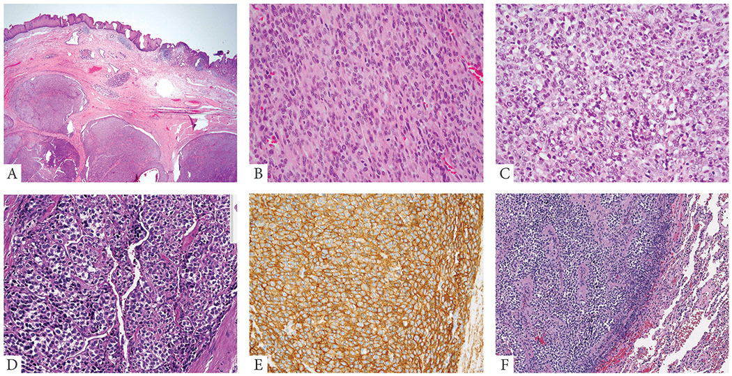
Figure 2: Morphologic spectrum of malignant GT with NOTCH gene fusions.

A. (67/M, esophageal, NOTCH2-MIR143) Low power image showing a multinodular lesion involving the submucosa of the gastro-esophageal junction. B. (77/F, small bowel, NOTCH2-MIR143) Malignant change in a GT in the form of sarcomatoid spindle cell areas with increased mitotic activity. C. (49/F, GE junction, NOTCH2 rearranged) Malignant GT in the form of undifferentiated round cell areas. D, E. (19/F, esophagus, NOTCH2-MIR143) Tumor shows a vague nested growth pattern, with epithelioid cells having sharply demarcated cell borders and variable clear cell change. Immunohistochemical stain for SMA (E) showing diffuse positivity in the tumor cells. F. (54/F, lung met, NOTCH2-MIR143) Metastatic GT to the lung with undifferentiated round cell morphology.
