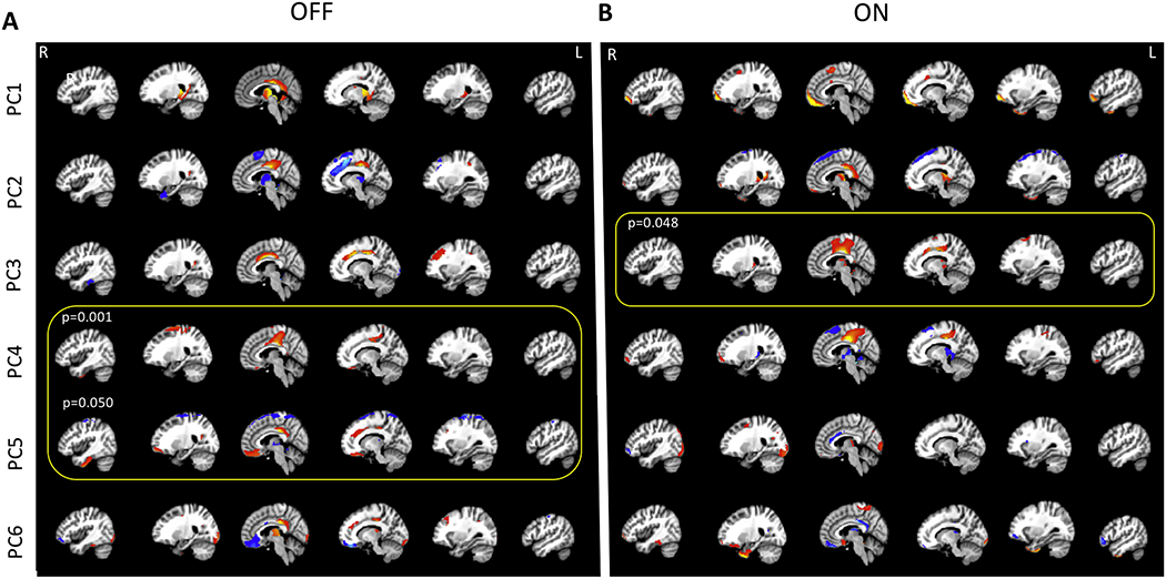
Figure 3.

(A) The first six PCs explaining about 50% of the variability in the data comparing control participants to PD subject OFF dopaminergic medication and (B) ON medication. In the OFF condition, PC1, explaining 15.2% of the variance, comprised of posterior cingulate, precuneus, thalamus, caudate, and cerebellum. PC2 explained 10.5% of the variance and included posterior cingulate, superior frontal gyrus, and the supplementary motor cortex. PC3 (explained variance 7.4%) included posterior cingulate, anterior cingulate, L middle frontal gyrus, while PC 4 (explained variance = 5.9%) comprised of posterior cingulate, postcentral gyrus, precentral gyrus, superior frontal gyrus, frontomedial cortex/subcallosal cortex, L occipital cortex. PC5 and PC6 explained 5.7 and 4.6% of the variance. PC5 included posterior cingulate, superior frontal gyrus, frontomedial cortex/subcallosal cortex, precentral gyrus and supplementary motor cortex, and PC6 included posterior cingulate, cuneus, bilateral temporo-occipital regions. In the ON state, PC1, explaining 15.8% of the variance, comprised of the frontal pole, supplementary motors cortex, and parahippocampal gyrus. PC2 explained 12.8% of the variance and included frontal pole, thalamus, parahippocampal gyrus, and the posterior cingulate. PC3 (explained variance 7.4%) included posterior cingulate, precentral gyrus, supplementary motor cortex, while PC 4 (explained variance = 5.7%) comprised of posterior cingulate, R frontal pole, and thalamus. PC5 and PC6 explained 5.0 and 4.3% of the variance. PC5 included Thalamus and R occipital cortex. PC6 included postcentral gyrus, precentral gyrus, frontomedial cortex/subcallosal cortex, and temporo-occipital regions.
