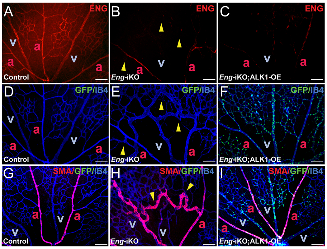
Figure 4. ALK1-OE restores aberrant SMC coverage observed in Eng-iKO retinal vasculature.

A-F, ENG (red) and IB4 (blue) fluorescence staining with retinas isolated from PN7 control (SclCreER-negative Eng-iKO, A and D), SclCreER;Eng-iKO (B and E), and SclCreER;Eng-iKO;ALK1-OE (C and F) mice. G-I. Double staining of IB4 (blue) and SMA (red) in PN7 retinas of controls (G), SclCreER;Eng-iKO (H), and SclCreER;Eng-iKO;ALK1-OE (I) mice. GFP reporter expression in IB4-positive retinal ECs in SclCreER;Eng-iKO;ALK1-OE (F and I). Yellow arrowheads indicate developed retinal AVMs. a, artery; v, vein. Scale bars: 100 μm.
