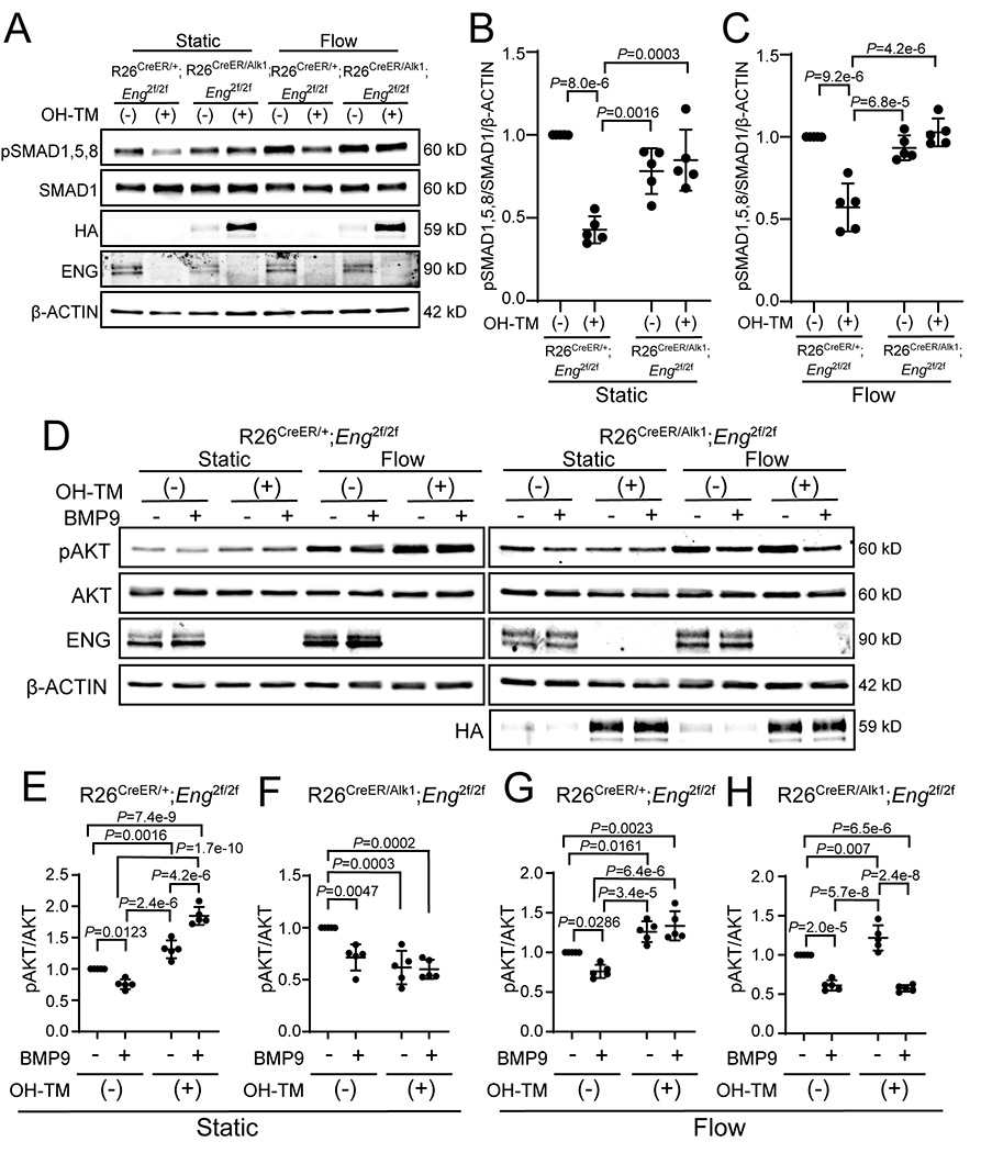
Figure 7. ALK1–OE leads to the rescue of disrupted SMAD and AKT signaling in Eng-iKO ECs.

A, Western blot analyses of phosphorylated SMAD1,5,8 (pSMAD1,5,8), ENG, and HA in pulmonary ECs from R26CreER;Eng-iKO or R26CreER;Eng-iKO;ALK1-OE. HA indicates transgenic ALK1 expression. One μM of 4-hydroxy-tamoxifen was treated to delete the Eng gene and to induce HA-ALK1 expression. BMP9 (0.5 ng/mL) was added to serum-starved pulmonary ECs for 45 min in the static or flow condition. B and C, Quantification of SMAD1,5,8 phosphorylation levels in static condition (B) and flow stimulation (C). pSMAD1,5,8 levels were normalized with total SMAD1 and β-ACTIN. n=5 per each group. Two-way ANOVA with Tukey’s correction. D, Western blot analyses of phosphorylated AKT (pAKT) and ENG in pulmonary ECs of R26CreER;Eng-iKO (left panel) or R26CreER;Eng-iKO;ALK1-OE (right panel). HA indicates ALK1 overexpression. One μM of OH-TM was used to delete the Eng gene. BMP9 (10 ng/mL) was treated for 90 min, followed by 30 min exposure to flow. E-H, Quantification of relative phosphor-AKT levels normalized with total AKT in static condition (E and F) and flow stimulation (G and H). n=5 per each group. Two-way ANOVA with Tukey’s correction. All data are means ± SDs.
