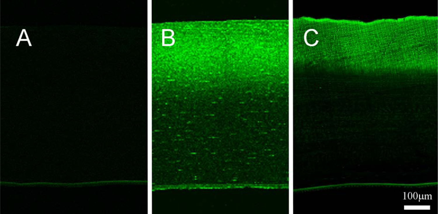
Figure 1: CAF Images.

(A) shows a representative CAF image of a control cornea with no crosslinking, (B) shows a cornea treated with UVA CXL, and (C) shows a cornea treated with NLO CXL using a Zeiss LSM 510 confocal microscope. CAF was detected in the 400–450 nm range, but images are shown in green to enhance contrast.
