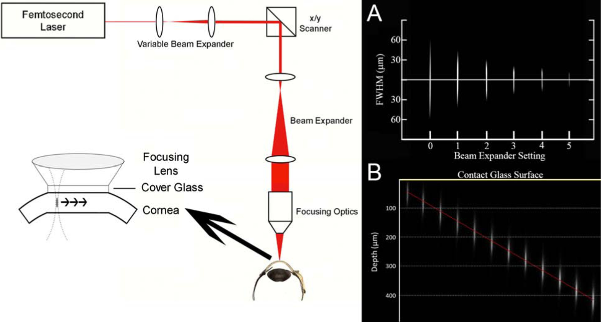
Figure 2: Delivery Device.

Schematic of designed delivery device with software controlled x, y scanners, variable beam expander, objective, and cone with contact glass. (A) Images taken of the two-photon focal volume within a tank of Rf solution as the focus is (A) changed in NA or (B) changed in depth beneath the contact glass.
