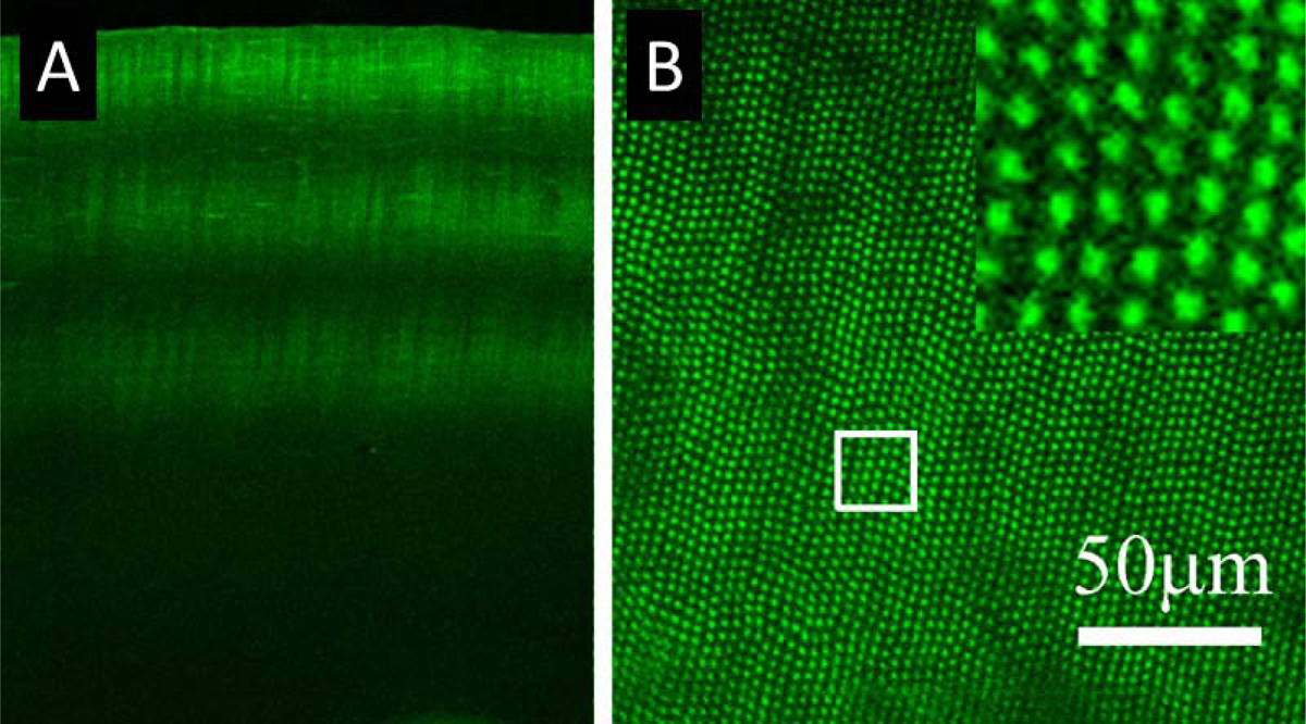
Figure 4: En Face CAF after a Single Amplified Pulse.

(A) A cross-sectional CAF image of a cornea treated with amplified FS pulses. (B) An enface CAF image of a cornea treated with single pulse NLO CXL as proof of concept for amplified pulse crosslinking. No pulses overlapped, and the non-crosslinked space between pulses is clearly visible as a drop in CAF intensity. CAF was detected in the 400–450 nm range, but images are shown in green to enhance contrast.
