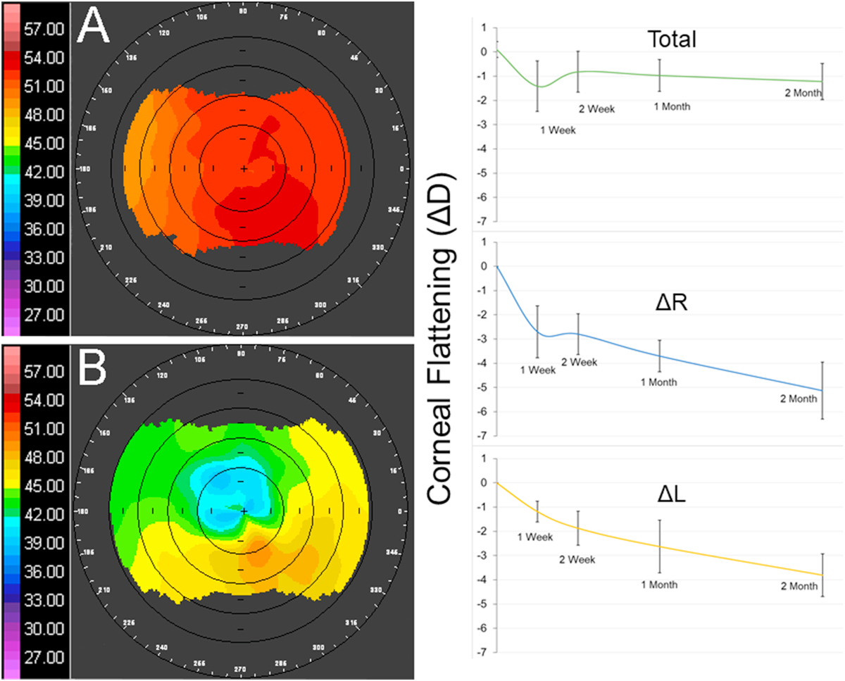
Figure 5: In Vivo Topography.

The corneal flattening of an in vivo rabbit cornea between baseline measurements (A) and two months post NLO CXL treatment (B) is localized to the region of treatment, a 4 mm circular region within the central cornea. The graphs indicate the total corneal flattening (ΔD), flattening within the treated right eye (ΔR), or flattening within the control left eye (ΔL).
