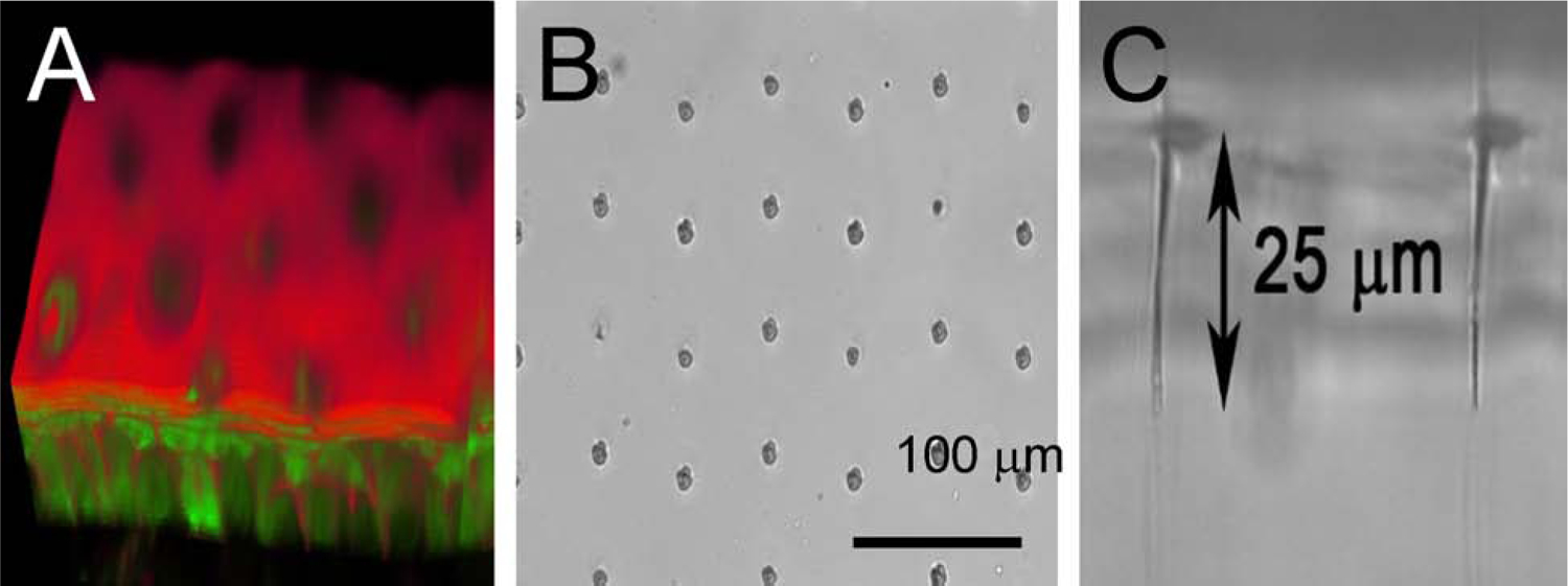
Figure 6: Channel Spacing in Silicone and Corneal Epithelium.

(A) A 3D reconstruction of a vibratome section of cornea from a microchannel treated corneal epithelium stained with Phalloidin and Propidium Iodide. Reconstruction based on a 3D stack of images taken with the laser scanning confocal microscope. (B and C) A grid of microchannels spaced 50 μm apart and 25 μm deep is shown is shown cut into a silicone sheet for demonstration.
