
Figure 7: Cellular Staining After Riboflavin Administration.
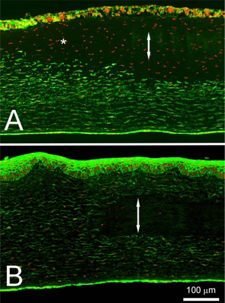
(A) shows a representative image of Phalloidin and Propidium Iodide stained cornea treated with transepithelial BAK riboflavin administration followed by NLO CXL and 24 hour culture. The cornea in (B) however was treated with microchannel riboflavin administration and NLO CXL. Double headed arrows represent the region of crosslinking treatment while the asterisk represents cellular damage outside of treatment region.