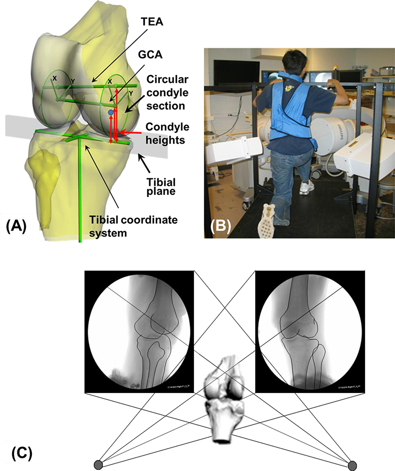
Fig. 1.
(A) The 3D knee joint model with tibial Cartesian coordinate system, the transepicondylar axis (TEA) and the geometrical center axis (GCA), the sagittal plane circular sections of the medial and lateral femoral condyles (including local coordinate systems on the circular sections). Measurements of femoral condyle heights with respect to the tibial cutting plane were also shown. (B) A subject performing a quasi-static single-legged lunge, which was captured the dual fluoroscopic system. (C) A virtual dual fluoroscopic system used for reproduction of the in vivo knee positions along the flexion path.

