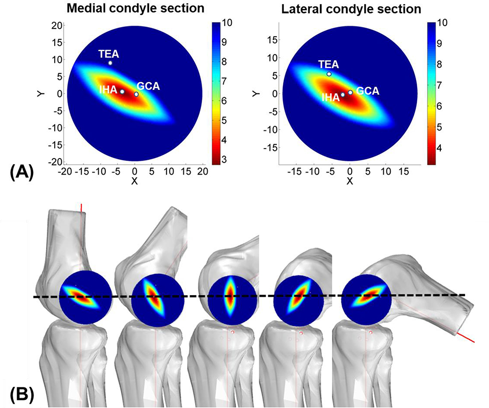
Fig. 2.
(A) Heat maps of the changes (mm) of the medial and lateral femoral condyle heights along the flexion path of the knee to illustrate the uniqueness of the IHA. The positions of the TEA, GCA and IHA on the condyle circular sections in full extension were marked. X and Y axes point to the posterior and proximal directions, respectively. (B) Diagrams showing the condyle height changes along the knee flexion path.

