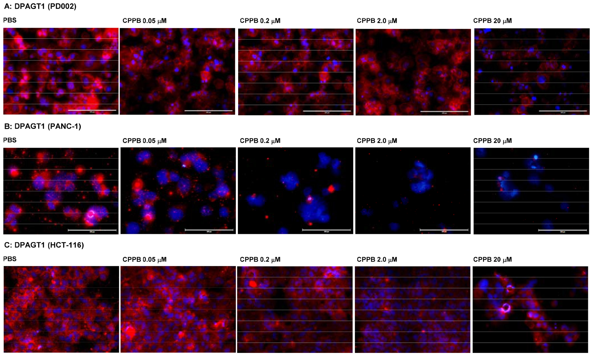
Figure 10.

Immunofluorescent staining: The DPAGT1 Expression Level in the Selected Cancer Cell Lines (PD002 and PANC-1) and a Colorectal Adenocarcinoma (HCT-116) Treated with CPPB.a
aFluorescence microscopy images at 20x. The cells (1 ×10⁵–6) were treated with CPPB (0.05, 0.2, 2.0, and 20 μM) or PBS for 72h. The cells were treated with DPAGT1 polyclonal antibody (Invitrogen, PA5–72704), followed by secondary antibody, Donkey anti-rabbit IgG (H+L), Alexa Fluor™ 555 (red) (Invitrogen). DAPI (4′,6-diamidino-2-phenylindole), a blue fluorescent DNA dye, was used to mark the nucleus.
