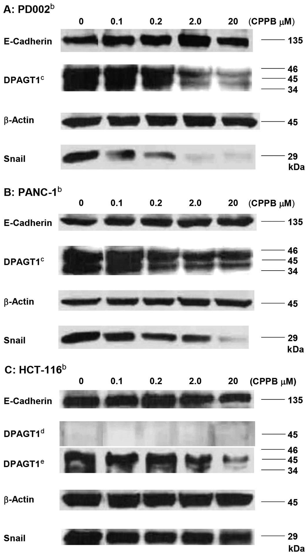
Figure 9.

Western blot Analyses of DPAGT1, Snail, and E-Cadherin in PD002, PANC-1, and HCT-116 treated with CPPB.a
aThe relative expression level was quantified by using Image Studio™ Lite quantification software (n = 3, p<0.001, see SI).; bAll cell lysates were prepared to be 1.5 mg total protein/mL by 15,200 xg, 30min at 4 °C unless indicated. CAt least three isoforms of DPAGT1 were detected.; dDPAGT1 was not detectable at 30 μL (1.5 mg total protein /mL).; eThe cell lysate was prepared by ultracentrifugation (130,000 xg for 1h at 4 °C). 30 μL of the lysate was analyzed.
