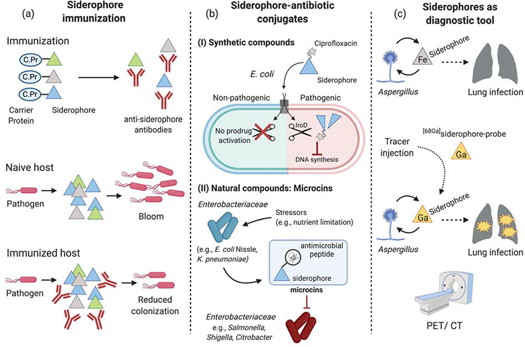
Figure 4. Siderophores as therapeutic targets and diagnostic tools.
(a) During infection, various siderophores are secreted by pathogens to thrive in the host despite host-mediated iron limitation. In experimental models, siderophores have been targeted for vaccination by linkage to a carrier protein in order to generate anti-siderophore antibodies. Whereas pathogens (e.g., Salmonella, UPEC) were able to successfully replicate in naïve hosts, siderophore-based immunization reduced their colonization levels. (b) Conjugation of antimicrobial peptides or antibiotics to siderophores. (b.I) An enterobactin-ciprofloxacin conjugate is transported through a common siderophore receptor found in the outer membrane of all E. coli strains. Once in the cytoplasm, the IroD esterase, which is mainly found in pathogenic E. coli strains, processes the enterobactin-ciprofloxacin prodrug and thereby enables ciprofloxacin-mediated inhibition of DNA synthesis. (b.II) Certain strains of Enterobacteriaceae, such as E. coli Nissle or K. pneumoniae, secrete microcins in response to nutrient (e.g., Fe) limitation. It is generally thought that the siderophore moiety enables these small antimicrobial peptides to better target susceptible strains of Enterobacteriaceae by utilizing their siderophore receptors for import. (c) Microbial siderophore uptake during infection can be leveraged for imaging applications. During lung infection, Aspergillus scavenges iron (Fe) via siderophores. Injection of siderophores radiolabeled with 68Ga in experimental Aspergillus infections enabled the visualization of the lung infection site by PET/CT.

