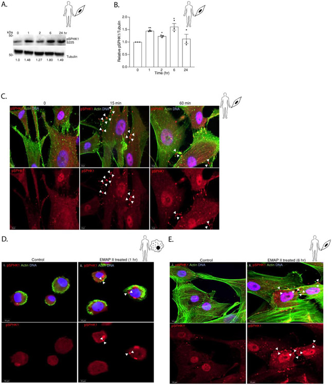
Figure 6. EMAP II induces bi-modal phosphorylation and plasma membrane localization of SPHK1.
(A) Representative western blot probed for pSPHK1 in whole cell lysates of hPASMC cells following EMAP II treatment for 0, 1, 2, 6 and 24 hours (B) quantitation of pSPHK1/ Tubulin. (C) Representative immunofluorescence images of EMAP II treated primary hPASMC at 0, 15 min and 60 min. (D) Representative immunofluorescence images of vehicle (i) EMAP II (ii) treated THP1 human macrophages at 1 hour. (E) Representative immunofluorescence images of vehicle (i) EMAP II (ii) treated primary hPASMC at 6 hours. (red= pSPHK1, green= actin and blue= DNA and arrows indicate the membrane localization of pSPHK1), scale bar is 10 μm. Results are shown as means ± SEM. n=3, *p<0.05 using unpaired t-test.

