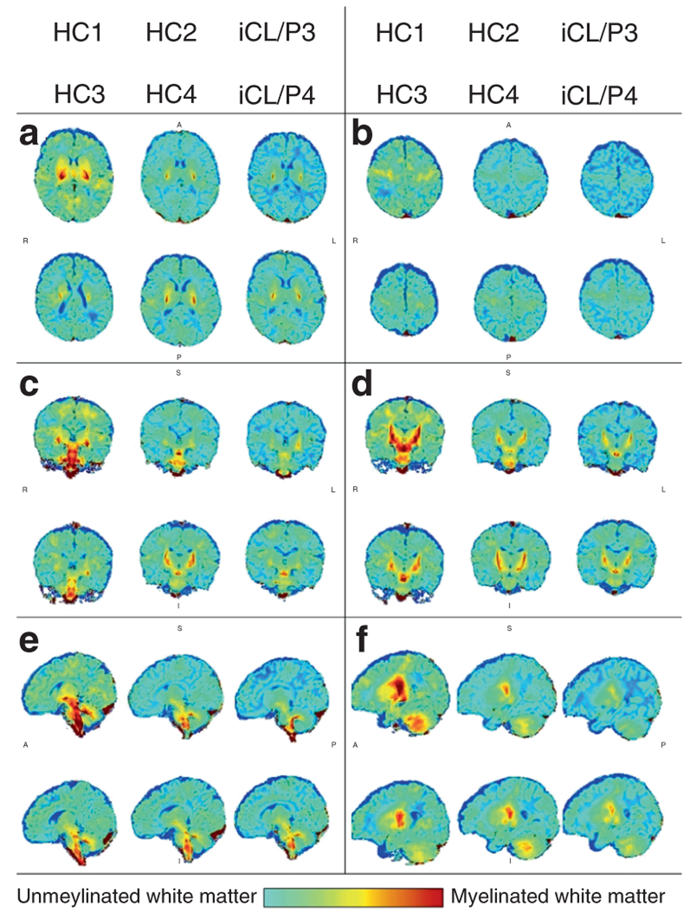
Fig. 2. Map of unmyelinated and myelinated white matter for individual participants.

The anterior commissure (AC; 100,133,81) is the center reference point; positive values are superior, right, and anterior to the AC. The following images present individual maps with sagittal slices a (x,y,5) and b (x,y,24); coronal slices b (x,−18,z) and c (x,−22,z); and axial slices d (24,y,z) and (36,y,z).
