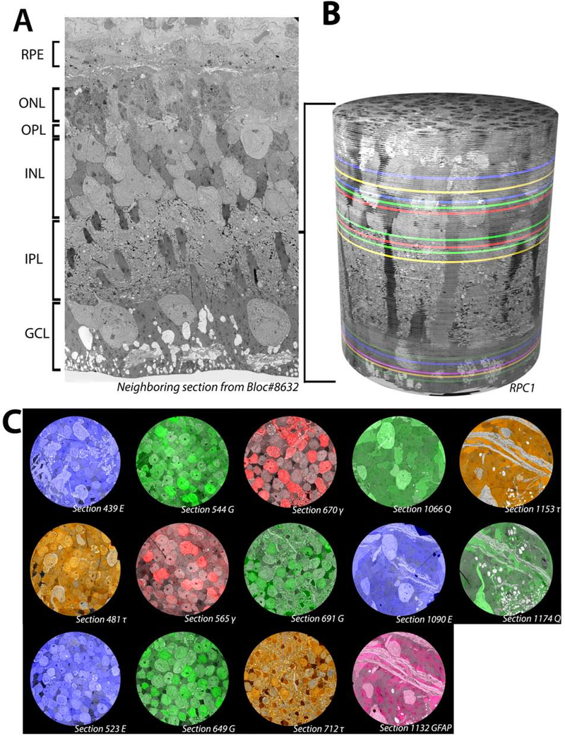
Figure 1.
Overview of RPC1. (A) Vertical section through tissue directly adjacent to the tissue processed for the RPC1 volume. (B) 3D composite volume of RPC1. Pseudocolored sections illustrate the locations of CMP sections for small molecules indicated in 1C. (C) Overlaid CMP sections from RPC1 on their adjacent TEM sections.

