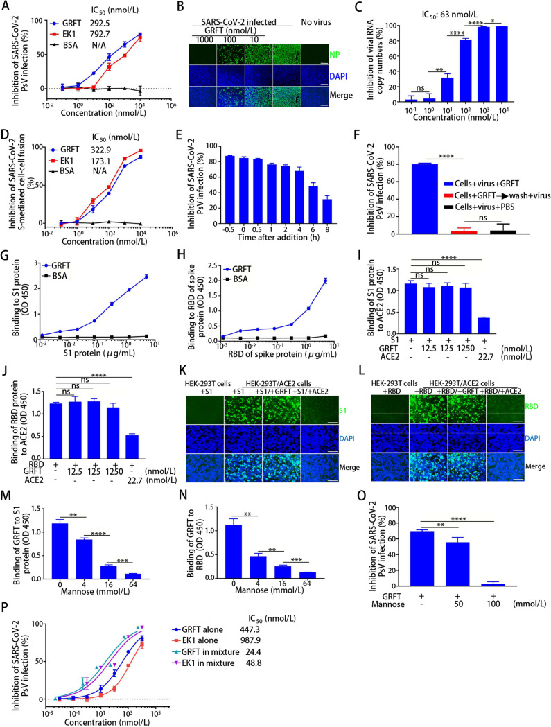
Fig. 1.
Inhibition of GRFT on SARS-CoV-2 infection and its mechanism of action. A–D GRFT inhibited infection of pseudotyped SARS-CoV-2 in HuH-7 cells measured by luciferase assay (A), and live SARS-CoV-2 (100 TCID50) in Vero-E6 cells measured by immunofluorescence assay (B), and qRT-PCR (C), as well as SARS-CoV-2 S-mediated cell–cell fusion in HuH-7 cells measured by immunofluorescence assay. Scale bars, 200 μm. (D). E–H Identification of the target site of GRFT. Time-of-addition assay (E). GRFT was added to HuH-7 cells 0.5 h before, during (0 h) and 0.5, 1, 2, 4, 6, and 8 h after SARS-CoV-2 infection. After 12 h incubation, culture supernatant containing GRFT was replaced by fresh medium, followed by culture for 48 h. The inhibitory activity of GRFT on SARS-CoV-2 pseudovirus infection was assessed by luciferase assay. Time-of-removal assay (F). Binding of GRFT to S1 subunit (G) and RBD (H) as measured by ELISA. I–L The effect of GRFT on the binding of S1 subunit (I) and RBD (J) to ACE2 as measured by ELISA, and on the binding of S1 subunit (K) and RBD (L) to HEK-293 T/ACE2 cells as measured by immunofluorescence assay. Scale bars, 200 μm. M–O Effect of mannose on GRFT binding to S1 subunit (M) and RBD (N) measured by ELISA, and on GRFT-mediated inhibition of SARS-CoV-2 pseudovirus infection (O). P Inhibition of SARS-CoV-2 pseudovirus infection by GRFT alone, EK1 alone, and their combination. Statistical analysis was performed and calculated by GraphPad Prism 5.0. The CalcuSyn program (kindly provided by Dr. T. C. Chou) was used to calculate the synergistic effect of combinations using the median effect equation (Chou TC 2006). *P<0.05; **P<0.01; ***P<0.001; ****P<0.0001; ns, not significant. See supplementary materials for the details of all experiment

