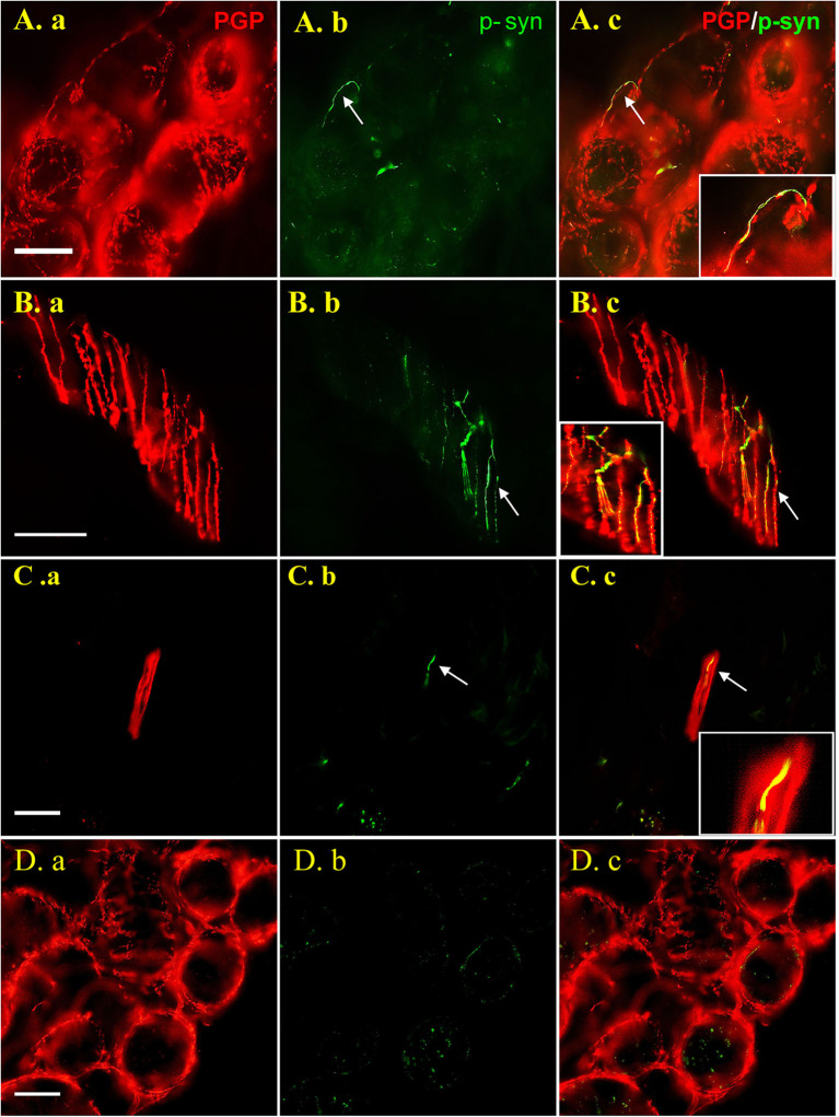
Figure 1.
Images are shown for PD patients (A–C) and healthy control subjects (D). Double labeling with anti-p-syn (green) and anti-PGP9.5 (red) were shown in 50 μm thick sections (A,B) and 15 μm sections (C). P-syn deposits (arrows) can be seen in nerve fibers innervating sweat gland (A.b), arrector pili muscle (B.b) and dermal nerve bundle (C.b) labeling by PGP9.5 (A.a, B.a, C.a). No p-syn deposition (D.b) was observed in sweat gland of the control (D.a). Figures were merged and magnified in A.c, B.c, C.c, and D.c. Bar = 50 μm. White arrow indicate p-syn positive deposition or PGP9.5/p-syn double positive position. PGP9.5: protein gene product 9.5; p-syn: phosphorylated alpha synuclein.

