Figure 5.
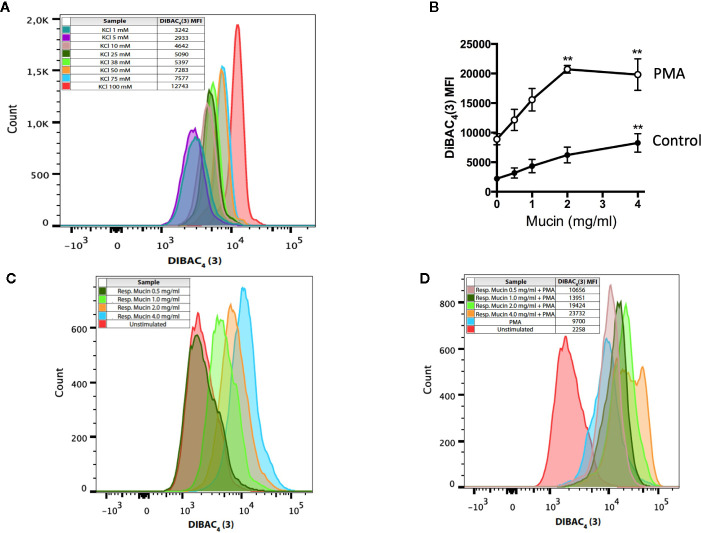
Respiratory mucins promote and potentiate PMA-induced neutrophil membrane depolarization. (A) Neutrophils were exposed to graded K+ concentrations. Membrane depolarization is indicated by the shift in the intensity of DiBAC4(3) fluorescence. Graded potassium solutions were made by increasing the KCl concentration in the solution. For graded K+ solutions, the KCl concentration was set to 1, 5, 10, 25, 38, 50, 75, and 100 mM. (B) Human neutrophils were incubated in the presence of respiratory mucins (•, 0.5–4 mg/ml) or stimulated with 200 nM PMA in the presence of respiratory mucins (<). After a 20-min incubation, the cells were stained with 100 nM DiBAC4(3). The transmembrane potential was determined by flow cytometry as described in Materials and Methods and is presented as mean fluorescence intensity (MFI) of the DiBAC4(3) probe. Means ± s.e.m. of four independent experiments are shown. (C) Representative flow cytometric histograms showing a concentration-dependent increase in DiBAC4(3) fluorescence in resting neutrophils incubated with mucins, indicating plasma membrane depolarization. (D) Exposure of neutrophils to increasing concentrations of respiratory mucins potentiated membrane depolarization in PMA-stimulated neutrophils. The data are representative of four separate experiments in four donors. (**p < 0.01 vs. no mucin).

