FIG 8.
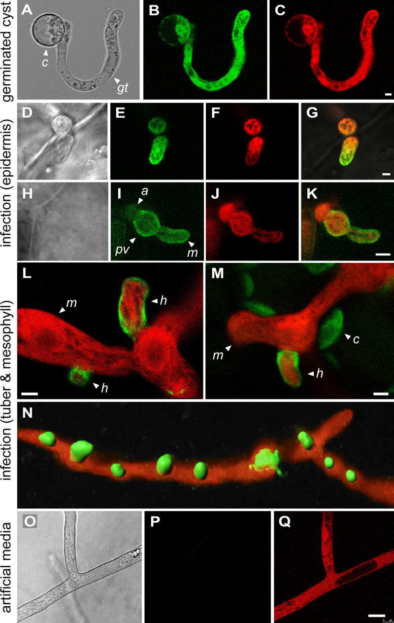
Localization of invertase expressed from native promoter. (A to C) Germinating zoospore cyst from strain expressing invertase fused with GFP and a separate gene encoding cytoplasmic tdTomato. Panels show bright-field, green, and red channels, from left to right. The cyst (c) and germ tube (gt) are marked. (D to G) Strain coexpressing invertase-GFP and cytoplasmic tdTomato in tomato leaf epidermal cell. The appressorium (a) and a young intracellular mycelium (m) are marked. Panels show bright-field, green, red, and merged images, from left to right. (H to K) Similar to panels D to G but at a later stage of growth. An appressorium (a), intracellular primary vesicle (pv), and mycelium (m) are marked. (L) Strain expressing invertase-GFP and cytoplasmic tdTomato in the medulla of an infected potato tuber. Emerging from the mycelium and penetrating host cells are several haustoria (h). (M) Same as panel L but showing mesophyll of a tomato leaf. The green channel detects both a GFP signal surrounding the haustorium (h) and autofluorescing chloroplasts (c). (N) Similar tissue as in panels L and M but showing a z-stack reconstruction of haustoria formed in the plant using Imaris software. (O to Q) Same strain as panel N but illustrating growth in rye-sucrose medium, showing an absence of invertase-GFP expression. Panels show bright-field, green, and red channels, from left to right. Each labeling pattern was observed in a minimum of three transformants. The images were obtained with PITG_14238, but similar results were obtained with PITG_14237. Bars, 2.5 μm.

